AI Triage for Chest X-Ray and CT Scans
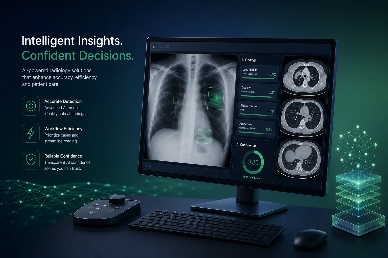
A radiology decision-support system that analyses chest X-rays and CT scans as they arrive, highlights suspected findings and moves urgent studies up the reading worklist, with radiologists making every diagnosis.
Project overview
We built an AI triage and decision-support system for a diagnostic imaging provider that reads chest X-rays and CT scans for hospitals and clinics. As studies arrive, the system analyses them, marks regions with suspected abnormalities and raises the priority of studies that may be urgent, so radiologists see the most time-critical cases first.
The engagement covered clinical discovery, data de-identification, annotation tooling, model development with PyTorch and MONAI, a FastAPI inference service, and integration with the provider's picture archiving (PACS) and reporting worklist. It is part of our healthcare software development practice and our work across the healthcare industry.
The client and the challenge
Studies were read in order of arrival. A chest X-ray showing a possible collapsed lung could sit behind dozens of routine follow-ups, especially overnight when fewer radiologists were on shift. The provider had no automatic way to tell urgent studies apart until a radiologist opened them.
The client wanted a system that fitted into existing PACS and reporting tools rather than another screen, supported radiologists instead of producing diagnoses, handled images from many scanner makes and hospitals, and treated patient data with strict privacy, consent and audit controls throughout development and operation.
Solution architecture & delivery
When a study is stored in PACS, a DICOM routing rule sends a copy to the inference gateway. The gateway checks modality, body part and series description, discards studies outside the intended use, and passes chest X-rays and chest CT series to the FastAPI inference service. Patient identifiers are replaced with an internal key before images reach the models, and the mapping stays inside the provider's network.
For chest X-rays, a convolutional classification network with localisation outputs, trained in PyTorch, predicts the likelihood of findings such as pneumothorax, pleural effusion, consolidation and nodules, and produces heatmaps showing where it is looking. For CT, MONAI pipelines handle volume loading, resampling to a consistent voxel spacing and intensity windowing, then a 3D segmentation network outlines suspected nodules and other lesions slice by slice. Training data came from de-identified historical studies used under the provider's data governance approval, annotated by radiologists in a labelling tool we set up, with a second reader resolving disagreements.
Results return to the clinical systems in standard formats: a structured report and overlay series stored back in PACS, and a priority flag sent to the reporting worklist so suspected urgent findings move up. The radiologist opens the study as usual, toggles the overlay, and confirms or rejects each finding; the AI never writes into the final report.
Because scanners and patient populations differ between hospitals, we validated performance per site and per scanner vendor before enabling the service there, and monitor input characteristics and radiologist agreement over time to catch drift. All access is logged, data is encrypted in transit and at rest, and the hosting follows the controls described on our security and compliance page. A related project is our hospital management suite, which covers the wider clinical workflow.
Key features we built
- Automatic analysis of chest X-rays and chest CT as studies arrive
- Suspected findings flagged with heatmaps and lesion outlines
- Worklist prioritisation for potentially urgent studies
- Overlay series and structured results stored back in PACS
- Radiologist confirm or reject controls on every finding
- De-identification before images reach the models
- Per-site and per-scanner validation before activation
- Drift monitoring and full audit logging
How we delivered it
- Clinical discovery. Workshops with radiologists and IT to agree the intended use, target findings, worklist behaviour and how results should appear in their existing viewer.
- Data governance and annotation. De-identification of historical studies under the provider's approval process, setup of the annotation tool and double-reading of labels by radiologists.
- Model development. Two-week sprints training and evaluating X-ray and CT models on held-out studies from different hospitals and scanner makes, with error review sessions alongside radiologists.
- Silent evaluation. The system ran on live studies without showing results, so its outputs could be compared with final radiologist reports before anything was visible to readers.
- Go-live and support. Site-by-site activation, monitoring and periodic revalidation under a support agreement, with release checks from our CI/CD testing pipeline.
Technology choices
- PyTorch — the deep learning framework for training and serving the chest X-ray classification and localisation network and the CT segmentation model.
- MONAI — medical imaging building blocks for DICOM and volume loading, spacing and intensity transforms, 3D networks and sliding-window inference on CT.
- FastAPI — an asynchronous inference API behind the DICOM gateway, with typed result schemas that map cleanly to structured reports and worklist updates.
Outcomes
- Studies with suspected urgent findings are moved ahead in the worklist instead of waiting in arrival order
- Radiologists review AI overlays inside their usual viewer rather than a separate application
- Every finding is confirmed or rejected by a radiologist, and the final report remains theirs
- Patient identifiers never reach the models, and all access is logged
- Performance is checked per hospital and scanner before activation and monitored afterwards
FAQ
Does the AI make diagnoses?
No. It is decision support. It flags suspected findings and changes worklist priority, but a radiologist reviews every study and writes the report. The system never adds text to the final report.
How is patient privacy protected?
Identifiers are replaced before images reach the models, training used de-identified studies under the provider's data governance approval, data is encrypted in transit and at rest, and every access is audited.
Do regulatory requirements apply to medical imaging AI?
Clinical AI is often regulated as a medical device depending on intended use and market. We designed documentation, validation records and change control so the provider could pursue the approvals relevant to them; we do not claim certification on their behalf.
How much does a medical imaging AI system cost?
It depends on the modalities and findings covered, annotation effort, PACS integration and validation scope. Our app development cost calculator gives an indicative range for the software side, and we estimate modelling and validation after discovery.
AI Triage for Chest X-Ray and CT Scans screens
Build your next healthcare product with Next Olive
Share your requirements — we will propose scope, timeline and stack within one business day.